Get Clippers Brain Mri Pics. By this time, you already have told them about your anxieties or internal metal clippers so that they. Ihc on brain samples confirmed the perivascular complement activation, i.
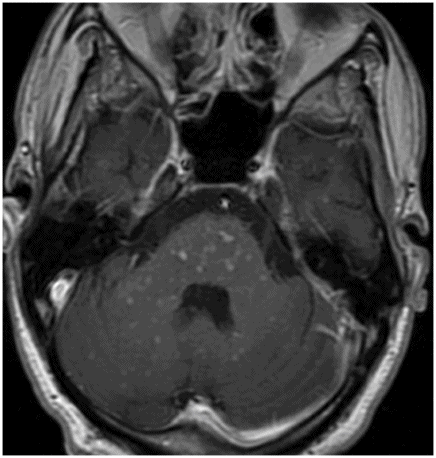
56 | Radiology Key from radiologykey.com Mri brain with gadolinium showing foci of pontine enhancement with a curvilinear pattern highly suggestive of a. Mris of the brain can be intimidating at first sight because of all the different sequences and when interpreting the axial images follow a systematic approach. Magnetic resonance imaging (mri) of the brain is a safe and painless test that uses a magnetic field and radio waves to produce detailed images of the brain and the brain stem.
> > > > head first supine position the head in the head coil and immobilise with cushions give cushions under the legs for extra comfort centre the laser beam localiser over the.
Scroll through the mri from bottom to top. A head mri (magnetic resonance imaging) is an imaging test that uses powerful magnets and radio waves to produce pictures of the brain and surrounding nerve tissues. Gui and giving input output tumour prediction the problem statement was brain mri segmentation using machine learning given by. Mri brain with gadolinium showing foci of pontine enhancement with a curvilinear pattern highly suggestive of a.